
Lymphedema Causes, Pathophysiology, Diagnosis and Therapy
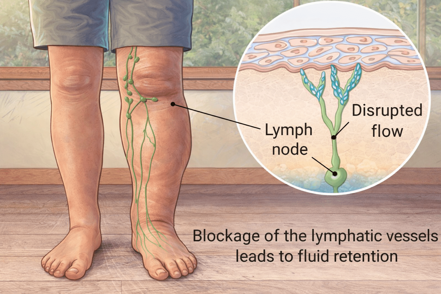
Lymphedema Guide Lymphedema: Causes, Pathophysiology, Diagnosis and Therapy Lymphedema is not just simple swelling. It is a chronic and progressive disease of the lymphatic system that requires early diagnosis, consistent therapy and lifelong management. In this article, we explain what causes lymphedema, how it progresses, how it is diagnosed and which therapies can help slow or stabilise the condition. By Ynone Team • Medical Education • 12 min read In this article What lymphedema is Etiology and classification Physiology of the lymphatic system Pathophysiology of lymphedema Clinical staging Diagnostic principles Complex Physical Decongestive Therapy Compression therapy Infections and skin care Selenium and immune support Patient education and long-term management Sources and references What lymphedema is Lymphedema is a chronic and progressive disease of the lymphatic system. It results from an insufficiency of lymph transport capacity and is characterised by the accumulation of protein-rich interstitial fluid. This accumulation leads to persistent tissue swelling, chronic inflammation, fibrosis and an increased risk of infections. Unlike transient or venous edema, lymphedema is not self-limiting and does not resolve spontaneously. Once established, it requires lifelong management. Clinically, lymphedema most frequently affects the extremities, particularly the arms and legs. It may also involve the trunk, head, neck or genital region. Over time, untreated or insufficiently treated lymphedema can lead to progressive tissue remodelling, skin changes and functional impairment, significantly reducing quality of life. Etiology and classification Lymphedema is classified as either primary or secondary, depending on its underlying cause. Primary lymphedema Primary lymphedema is caused by congenital malformations of the lymphatic system. These may include hypoplasia, aplasia or structural abnormalities of lymph vessels and lymph nodes. Primary forms typically present at birth, during puberty or in early adulthood and predominantly affect women. The lower extremities are most commonly involved, often unilaterally. The swelling usually begins distally and progresses proximally over time. Secondary lymphedema Secondary lymphedema develops as a result of acquired damage to an initially intact lymphatic system. In industrialised countries, the most common causes are oncological interventions, particularly surgical removal of lymph nodes and radiotherapy. Additional causes include trauma, burns, recurrent infections, inflammatory processes, malignant tumours compressing lymphatic pathways and, in some regions, parasitic infections such as filariasis. When no clear cause can be identified, the condition is described as idiopathic lymphedema. Physiology of the lymphatic system Under physiological conditions, blood circulation continuously delivers oxygen and nutrients to tissues while removing metabolic waste products. At the level of the capillaries, fluid is filtered into the interstitial space. Approximately ninety percent of this fluid is reabsorbed directly into the venous system. The remaining ten percent, amounting to six to ten litres per day, remains in the interstitial space and must be removed by the lymphatic system. This residual fluid, known as lymph, contains plasma proteins, lipids, immune cells, cellular debris, microorganisms and foreign particles. The lymphatic system begins as a network of open-ended lymph capillaries embedded in the connective tissue. Through pressure gradients, tissue movement and intrinsic vessel contractility, lymph is absorbed and transported through pre-collectors, collectors and lymph nodes before being returned to the venous circulation. Beyond fluid balance, the lymphatic system is a central component of immune defence. Lymph nodes function as filtration and immune activation sites, enabling the recognition and elimination of pathogens and foreign substances. Pathophysiology of lymphedema Lymphedema develops when the transport capacity of the lymphatic system is reduced below the required load. This may occur due to structural damage, functional impairment or both. As lymph transport becomes insufficient, protein-rich fluid accumulates in the interstitial space. The presence of proteins in the tissue initiates a chronic inflammatory response. Fibroblasts are activated, leading to progressive fibrosis, while adipose tissue deposition increases over time. These changes explain why lymphedema gradually becomes firmer, less compressible and increasingly resistant to treatment if not addressed early. Diuretics are ineffective because they do not remove proteins from the interstitial space and may even worsen tissue changes. Clinical staging Lymphedema progresses through several clinically defined stages. Latent stage In the latent stage, lymphatic damage is already present, but no visible swelling can be detected. Functional diagnostic methods may reveal impaired lymph transport. Reversible stage In the reversible stage, soft swelling is present and pitting can be induced by pressure. Elevation of the affected limb leads to a temporary reduction in swelling. Spontaneously irreversible stage As the disease progresses, tissue fibrosis increases, pitting becomes difficult or impossible, and swelling no longer resolves with elevation. Elephantiasis In the most advanced stage, known as elephantiasis, massive swelling, pronounced skin changes and severe functional limitations occur. Diagnostic principles The diagnosis of lymphedema is primarily clinical and is based on a detailed medical history and physical examination. Imaging techniques such as lymphoscintigraphy or indocyanine green lymphography may be used in unclear cases or for treatment planning. Ultrasound and magnetic resonance imaging are helpful in excluding differential diagnoses such as venous disease or tumours. Complex Physical Decongestive Therapy The internationally accepted gold standard for the treatment of lymphedema is Complex Physical Decongestive Therapy, also known as CPT, Complex Physical Therapy or KPE. CPT consists of four interdependent components: Manual lymph drainage Compression therapy Exercise therapy Meticulous skin care These elements must be applied in a coordinated and consistent manner to achieve effective and sustainable results. Manual lymph drainage Manual lymph drainage is a specialised therapeutic technique designed to stimulate lymph transport. A fundamental principle of effective MLD is that treatment must always begin centrally, at the level of the neck. This region contains the venous angles where lymph enters the bloodstream. Clearing central lymphatic pathways first creates a suction effect that allows lymph from peripheral regions to drain effectively. If central clearance is neglected, peripheral drainage techniques are largely ineffective. Incorrectly performed MLD provides no therapeutic benefit and represents a misuse of time and resources. To maintain its effect, MLD must always be combined with compression therapy. Compression therapy Compression therapy is the most decisive and effective component of lymphedema treatment. By increasing tissue pressure, compression reduces capillary filtration, enhances lymph uptake, redistributes
Lymphedema Causes, Pathophysiology, Diagnosis and Therapy Read Post »